1Zorya I
A proton-driven ZorA5B2 motor; its tail recruits ZorC and ZorD to destroy phage DNA.
How does ZorAB sense the phage at the cell envelope?
Bacteria encode a diverse arsenal of anti-phage defence systems. Combining microfluidics, quantitative live-cell imaging and bacterial genetics, we resolve how these systems sense infection, how they are coordinated in time and space within single cells, and how they act together to protect the population.
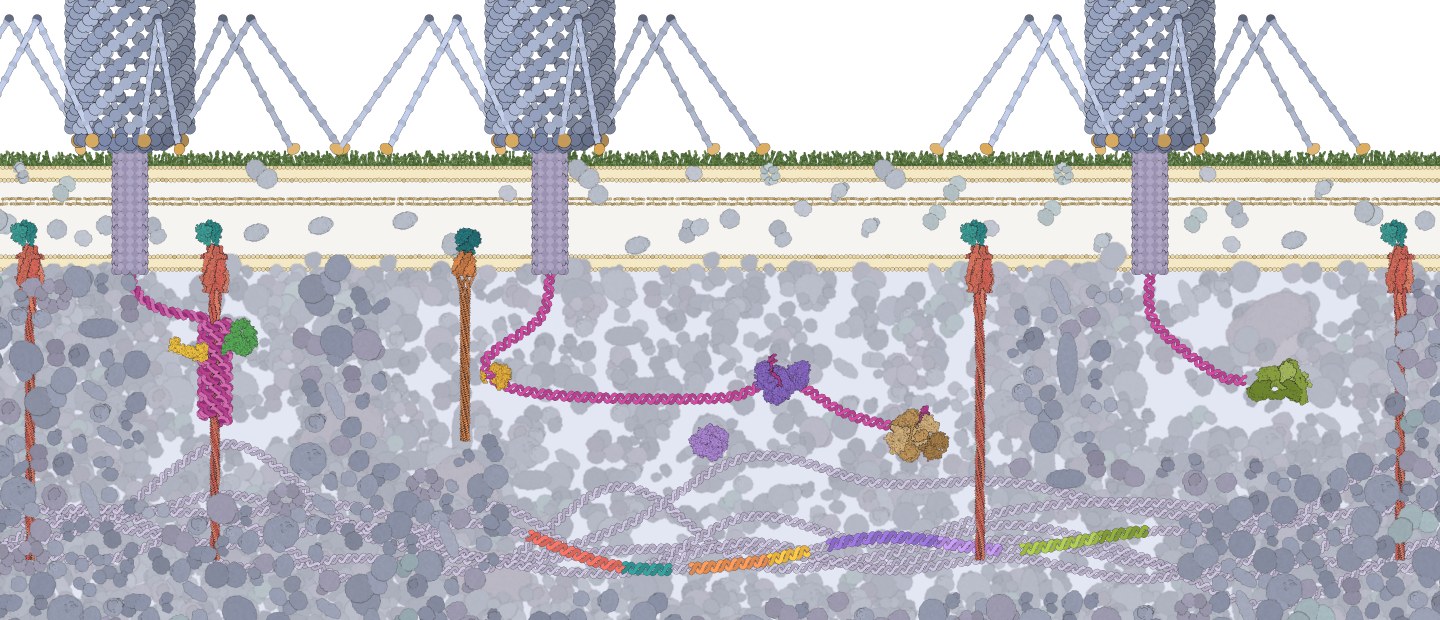
Zorya at work: phage infection deforms the cell envelope, so the ZorB2 domains can reach the peptidoglycan (how exactly the gap closes is not yet known). Protons then flow through the bound ZorB and drive the rotation of ZorA, whose tail reels in the phage DNA and ZorC and ZorD destroy it. Model based on Hu, Popp et al. 2025, Nature; DNA winding by the tail is a proposed mechanism.
Bacteria are under constant attack by phages and defend themselves with a remarkable diversity of immune systems. Most species carry several of them, often clustered in defence islands. How these modules are coordinated in time and space, and how they are embedded in the wider physiology of the cell, remains largely unknown.
Defence systems differ in how they sense infection, where they localise in the cell and when they act, and some confer protection only in combination. The figure maps the systems we study within a single cell, together with current mechanistic knowledge and the open questions we address.
 123456
Structures: ZorA5B2 8QYD and 8VVI, ZorC 8R68, ZorD 8QY7, ZorE AlphaFold P0DW03, DruE 9TU7, DruH 9TUD, RecBCD 1W36, LmuACB 9HQU. Phages, DNA, membranes and cytoplasm are schematic.
1Zorya I
A proton-driven ZorA5B2 motor; its tail recruits ZorC and ZorD to destroy phage DNA.
How does ZorAB sense the phage at the cell envelope?
2Zorya II
The same 5:2 motor core, paired with the nickase ZorE.
How is ZorE brought to the phage DNA?
3Druantia III
DruE recognises single-stranded phage DNA and unwinds it directionally.
Why is it so much stronger together with Zorya II?
4RecBCD
The cell's own DNA-end processing machine.
A shared hub that connects defence systems?
5Lamassu
Senses phage DNA ends and releases the nuclease LmuA: abortive infection.
When does the cell sacrifice itself instead of defending?
6Defence island
Several systems encoded side by side on the chromosome.
How are they coordinated in time and space in single cells?
We follow defence in single living cells and connect what we see to the structures of the defence complexes.
Reporter fusion in time-lapse microscopy over two hours.
We combine or remove defence systems in E. coli, build fluorescent reporters and label phages to follow infection dynamics.
Animation: labelled phages reach cells in a mother machine (with Dietrich Kohlheyer, FZ Jülich). Right: real channels with labelled phages.
Microfluidic mother machines keep thousands of single cells under constant conditions while phages arrive. Live-cell and TIRF microscopy show where and when each defence system acts.
ZorA5B2 (PDB 8QYD) between the inner membrane and the peptidoglycan.
We aim to unravel the mechanistic details of anti-phage defence systems: with structural biologists we determine the architecture of defence complexes and test proposed mechanisms in living cells.
Hu, Popp et al. 2025, Nature · Himpich et al. 2026, Cell Reports
BactoMate: segmentation, foci, cell outlines and tracking.
BactoMate, our open-source platform, turns microscopy into single-cell data: segmentation, foci, tracking and lineages, in one workflow.

Einstein Independent Researcher and head of the Molecular Infection Microbiology group at the Institute of Biology, Humboldt-Universität zu Berlin. The group studies how anti-phage defence systems act together in single bacterial cells.


Nikos Moustakas
PhD student, since 2026
Coordinated immunity in a single cell
Valentin Schmitt
B. Sc. student, since 2026
Mechanism of Druantia immunity
PhD students and postdocs
We welcome PhD students and postdocs who want to apply for their own funding to work with us. We support fellowship applications, for example to the Alexander von Humboldt Foundation, Marie Skłodowska-Curie Actions, EMBO, DAAD or the Boehringer Ingelheim Fonds.
B. Sc. and M. Sc. theses
We offer thesis projects in microbiology and microscopy data analysis. Send us a short statement of interest and your CV, together with a short project idea or the methods you would most like to work with.
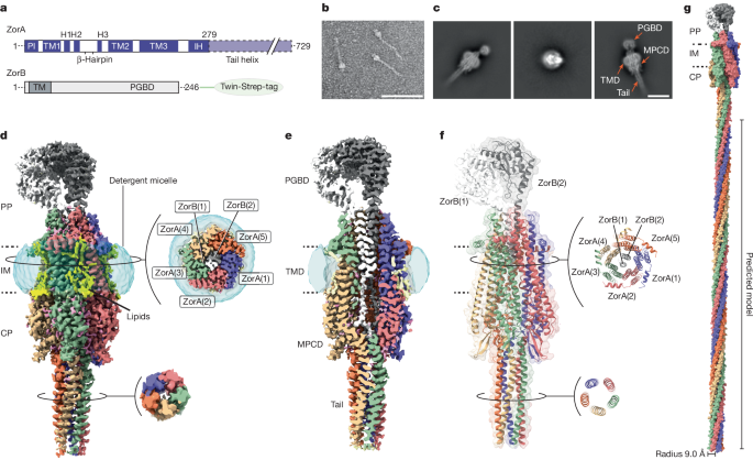 Nature 2025Structure and mechanism of the Zorya anti-phage defence systemZorAB is a 5:2 proton-driven rotary motor with a ~70 nm cytoplasmic tail that recruits nuclease effectors after phage sensing.Hu, Popp et al.
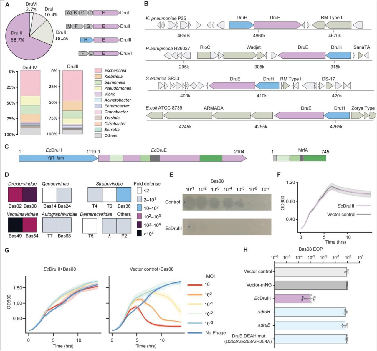 Cell Reports 2026A compact Druantia defense clears phage infections via ssDNA recognition and directional duplex unwindingDruantia recognizes phage ssDNA and drives directional duplex unwinding to clear the infection in a compact anti-phage system.Himpich et al.
 bioRxiv 2026BactoMate: an integrated platform for reproducible bacterial microscopy analysisAn end-to-end toolkit for conversion, segmentation, tracking, lineage QC and reporting of bacterial microscopy datasets.Hallenga et al.
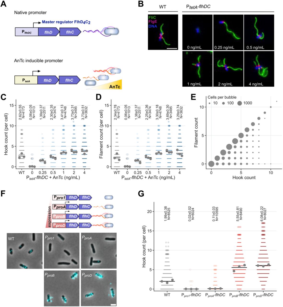 PNAS 2025Optimization of bacterial flagellum growth for efficient motility and energy consumptionA critical filament-length threshold balances swimming performance against the energetic cost of building the flagellum.Halte et al.
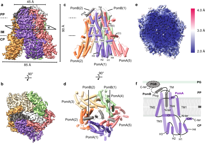 Nature Communications 2023Ion selectivity and rotor coupling of the Vibrio flagellar sodium-driven stator unitCryo-EM reveals how the sodium-driven MotPS stator selects ions and couples rotation to the flagellar rotor.Hu et al.
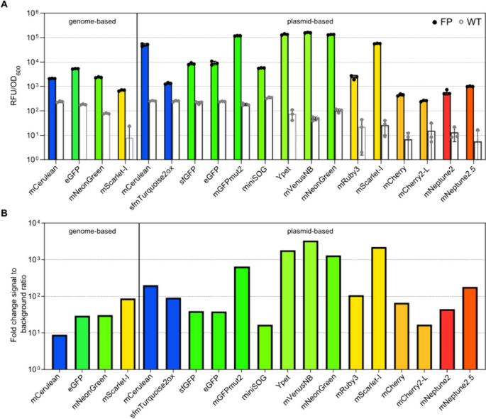 J. Biol. Eng. 2024Fluorescent tools for the standardized work in Gram-negative bacteriaA practical fluorescent toolkit for reproducible imaging and genetic work across Gram-negative bacteria.Delgadillo-Guevara et al.